Description
Transcription
Panel labels (italic white letters on black or image background, upper-left of each panel):
- a (top-left)
- b (top-right)
- c (bottom-left)
- d (bottom-right)
Instrument / acquisition data bars (white text on black strip along the bottom of each panel), left → right:
Panel a:
- BEC
- 10kV
- SS68
- 30Pa
- x50
- 500μm
- scale bar (white horizontal bar at far right of the data strip)
Panel b:
- BEC
- 10kV
- SS68
- 30Pa
- x100
- 100μm
- scale bar (white horizontal bar at far right of the data strip)
Panel c:
- BEC
- 10kV
- SS68
- 30Pa
- x190
- 100μm
- scale bar (white horizontal bar at far right of the data strip)
Panel d:
- BEC
- 10kV
- SS68
- 30Pa
- x75
- 200μm
- scale bar (white horizontal bar at far right of the data strip)
No figure caption, title, or legend text is printed on this cropped PNG itself (caption lives on the parent journal page, not in this image).
No coordinate grids, place names, or mineral-name annotations on the image faces.
Description
TYPE & PURPOSE
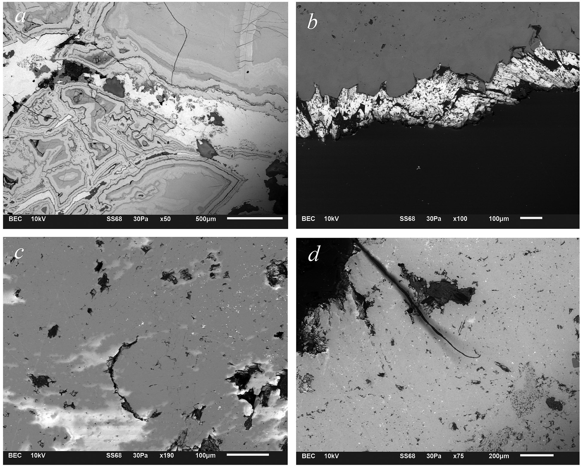
Four-panel backscattered-electron contrast (BEC) scanning electron microscope (SEM) micrograph collage of malachite (and associated phases) from the Rosen ore-field copper deposits (SE Bulgaria). The figure documents internal growth textures, banded/colloform microstructure, free surface morphology, and internal defects (voids, inclusions, fractures) of the malachite samples illustrated at hand-specimen scale in the companion Fig. 2 of the same paper. BEC mode encodes mean atomic number as brightness: denser / higher-Z phases appear lighter; lower-Z or porous/void regions appear darker.
FRAME, EXTENT, ORIENTATION
- Single composite figure: 2 × 2 rectangular grid of greyscale SEM panels.
- Arrangement:
- Top row: a (left) | b (right)
- Bottom row: c (left) | d (right)
- Each panel is a plan/section view of a polished or fractured mineral surface; no geographic north applies. Orientation is image-frame only (not a map).
- Thin white hairlines separate the four panels; each panel has its own black instrument-data strip along the bottom edge.
- Panel labels a–d sit in the upper-left corner of each frame in italic roman type.
SCALE
Each panel carries its own stated magnification and micrometre scale bar on the instrument strip:
| Panel | Magnification | Stated scale length |
|---|---|---|
| a | ×50 | 500 μm |
| b | ×100 | 100 μm |
| c | ×190 | 100 μm |
| d | ×75 | 200 μm |
Scale bars are thin white horizontal lines at the right end of each data strip. No common scale across panels; comparison between panels must use each panel’s own bar.
ACQUISITION PARAMETERS (common to all four panels)
Read from every data strip:
- Mode: BEC — backscattered electron contrast (compositional / Z-contrast imaging).
- Accelerating voltage: 10 kV.
- Spot / beam setting code: SS68 (instrument spot-size or scan-setting identifier as printed by the SEM UI).
- Chamber pressure: 30 Pa (low-vacuum / variable-pressure SEM mode, consistent with imaging uncoated or poorly conducting oxide/carbonate specimens without heavy charging).
- Magnification and scale length: panel-specific as tabulated above.
No detector branding, working-distance, or dwell-time values are printed beyond the fields above.
Panel (a) — Low-magnification BEC of banded / colloform malachite (×50, 500 μm bar)
What is shown. A polished (or planar) section through a concentrically and irregularly banded carbonate aggregate dominated by malachite growth banding. The field of view is filled almost entirely by the mineral mass; a dark (low-BSE) region occupies the lower-right corner of the frame and a smaller dark triangular embayment sits mid-right.
Texture and microstructure (observed):
- Concentric / colloform banding. Multiple nested, closed-to-open concentric bands occupy the left–central two-thirds of the frame. Bands form irregular quasi-elliptical to lobate rings with:
- alternate light-grey and medium-grey concentric shells,
- local dark-grey thin rims marking individual growth increments,
- thicker light-grey bands that include fine internal laminae.
- Core / nucleus region. Near the geometric centre-left of the concentrically banded body is a complex, crudely polygonal to amoeboid nuclear zone of mixed mid-grey and pale-grey material, with small dark voids and darker enclaves. This area is the apparent growth centre around which successive malachite bands accreted.
- Botryoidal / mamillary outer surface (sectioned). The lower and right margins of the banded body show scalloped, convex-outward growth fronts — classic colloform/botryoidal malachite geometry in cross-section.
- Fractures / cracks. Several thin dark linear cracks transect the banding:
- a prominent near-vertical hairline crack runs from the top edge roughly through the right side of the banded body into the middle of the frame;
- additional curved and branching cracks follow or cut band boundaries in the upper-left sector;
- a finer network of microcracks is visible in the lighter banded domains (desiccation or sample-preparation related, and/or natural brittle fracture).
- Inclusions / dark patches. Scattered irregular dark-grey to black patches (tens to >100 μm) occur inside and between bands — some appear to be voids/pores, others may be lower-Z inclusions or pulled-out phases. One larger irregular dark body sits mid-right above the thick outer light band.
- Outer host / matrix at upper and right margins. Beyond the main banded body, a smoother, more homogeneous mid-to-light grey field occupies the upper-right quadrant; it is crossed by fine cracks and shows fainter, nearly parallel laminar banding (sub-horizontal) rather than concentric rings — consistent with later, more planar crustiform overgrowth or with a different section orientation through the same aggregate.
- Porosity. Dark irregular voids and dissolution-looking embayments are concentrated along some band boundaries and in the nuclear area.
Grey levels (BEC implication). The dominant greys are mid-to-light, consistent with a copper carbonate (malachite, Cu₂CO₃(OH)₂) matrix. Darker zones are voids, cracks filled with mounting medium / vacuum, or lower-Z material. No extreme bright (high-Z sulfide or native-metal) phases dominate this panel — the image is essentially a single-mineral banded fabric with porosity.
Spatial scale of features. Individual growth bands are typically a few tens of micrometres thick; the whole concentrically banded body spans several millimetres across the field (the 500 μm bar occupies a small fraction of the frame width — roughly ~1/6–1/8 of the frame, so the FOV is on the order of ~3–4 mm across).
Panel (b) — Free surface / fracture morphology of malachite (×100, 100 μm bar)
What is shown. An edge-on or free-surface SEM view of a rugged, high-relief mineral surface (upper half of the frame is the sample; lower half is empty dark void / SEM background). The surface runs roughly horizontally as a ragged free edge across the middle of the panel, convex-upward in several places, with a nearly featureless mid-grey smooth face above the irregular margin.
Texture and microstructure (observed):
- Upper domain — smooth cleaved / polished face. The upper third to half of the panel is a planar, homogeneous mid-grey surface with only faint fine pits and rare small dark speckles. This is either a cleaved crystal face, a polished cut surface of the carbonate, or a flat fracture plane.
- Lower margin of the upper domain — rugged free surface. At the free edge the surface breaks into a highly irregular, jagged, continuously scalloped topography:
- numerous pointed, blade-like, and tongue-shaped projections hang downward into the dark field;
- re-entrants and deep notches separate the projections;
- surface of the projections is rough, white-to-light-grey in BEC, with granular/crystalline micro-texture rather than mirror-smooth.
- Microcrystalline / fibrous texture on the free surface. At this magnification the free-surface content of the projections resolves into a felted aggregate of tiny crystallites / fibrous needles / platy chips — morphology typical of malachite free surfaces and fractured botryoidal crusts. Individual crystallites are on the order of a few micrometres; fascicles of them give the bright granular look.
- Dark inclusions / voids in the free-surface zone. Several wedge- to lens-shaped black regions are nested within the bright free-surface material (mid-left, centre, right) — open pores, pulled-out grains, or deep recesses of the surface topography under BEC shadowing.
- Far-right edge. The free surface terminates against a darker, more massive block of material on the far right; a vertical-ish stepped contact is visible.
- Lower half of frame. Featureless near-black background (vacuum / no sample). A single tiny bright spot (debris or dust particle) floats in the dark field near centre.
Grey levels. Upper smooth face = mid-grey (solid malachite). Free-surface roughness appears preferentially bright white — common BEC/edge-highlight and topographic-contrast effect on high-relief carbonates at low voltage, and/or locally denser packing of crystallites. Black = void.
Scale of features. Free-surface relief amplitude (ridge tips to deep recesses) is tens of micrometres; the 100 μm bar spans a small strip near the right of the data bar. FOV width is on the order of ~1 mm.
Panel (c) — Internal fabric with voids and polycrystalline domains (×190, 100 μm bar)
What is shown. A higher-magnification BEC view of an interior polished (or slightly uneven) face of the malachite aggregate. The matrix is mid-grey and largely continuous; strewn through it are numerous dark irregular voids, darker soft blotches, and one prominent C-shaped / horseshoe dark vug near the lower-centre.
Texture and microstructure (observed):
- Host matrix. Continuous mid-grey phase occupying most of the field — the malachite groundmass. The surface is not perfectly flat: broad soft brightness gradients (mid-left brightening; lower-left bright wash) indicate mild topography or slight zoning in mean Z / density.
- Central–lower C-shaped vug. A large open void arcs as a dark C or incomplete ring opening toward the upper-right, roughly ~50–80 μm across the opening. Its walls are sharp against the matrix; thin bright lining films appear locally on the void wall (possibly residual coating, thin secondary film, or edge-brightening).
- Scattered irregular voids. Multiple smaller dark pores and angular voids are distributed across the field:
- upper-left cluster of angular dark chips/voids,
- upper-centre and upper-right flecks,
- mid-right larger angular dark body,
- lower-right cluster of irregular black patches,
- lower-left small flecks. Void shapes range from equant to elongated to jagged; sizes from a few μm to several tens of μm.
- Bright particulate / microcrystalline flecks. Numerous tiny bright white points and short streaks are sprinkled through the mid-grey matrix, especially in the upper half and around voids. These are consistent with either (a) higher-Z microminerals (e.g. residual Cu-sulfide or Fe-oxide micrograins), (b) topographic edge-highlights on micro-relief, or (c) charging artefacts on insulating microparticles. They are discrete, not a continuous second phase.
- Soft cloudy dark patches. Several diffuse medium-dark grey smudges (not sharp voids) occur mid-field and upper-mid — possibly sub-surface cavities under a thin roof, epoxy-filled pores, or compositional heterogeneity (less dense hydrated/altered zones).
- Faint linear microcracks. Thin short dark hairlines cross parts of the matrix (faint, low contrast) — microfractures.
Grey levels. Dominant mid-grey = malachite. Black = open voids. White flecks = higher-Z or edge effects. No large bright sulfide mass is present in this panel.
Scale of features. Highest-magnification panel of the set (×190). The 100 μm bar indicates FOV width roughly ~0.5 mm. The C-vug is a major feature at this scale; individual bright flecks are micrometre-scale.
Panel (d) — Internal fabric with fracture, inclusion cluster, and felted patches (×75, 200 μm bar)
What is shown. A mid-magnification BEC view of a broad interior face of the sample. Mid-grey continuous matrix dominates. A long, gently curved dark fracture/veinlet runs from upper-left toward lower-centre. Near the fracture midspan sits an irregular cluster of dark voids and mid-dark soft patches. Along the left margin a dark, fibrous/felted mass occupies a substantial sector.
Texture and microstructure (observed):
- Host matrix. Mostly homogeneous mid-to-light grey malachite mass with fine granular noise / microcrystalline texture under this magnification; densely sprinkled with very fine dark micropits across the whole field.
- Long curved fracture. A continuous dark linear feature — narrow, smoothly curved, concave to the right — extends from near the upper-left edge, through the mid-left, and trails off toward the lower mid-field. Locally it has appreciable width (filled crack or dissolution seam); near its mid-section it passes beside / through the dark inclusion cluster. Consistent with a brittle fracture, a healed joint, or a sheet-like partial dissolution seam.
- Central dark inclusion / void cluster. Immediately left to slightly above midframe (touching the fracture) is an irregular aggregate of:
- elongated and amoeboid black voids,
- medium-dark soft patches,
- and a roughly stellate/branching outline. Overall cluster diameter is on the order of a bit under 100 μm (visual relative to the 200 μm bar). Likely a compound pore space, a pulled-out inclusion nest, or a residual remnant of a replaced precursor phase.
- Left-margin felted / fibrous dark mass. On the far left edge (spilling top-left to mid-left) is a sizable dark-to-mid-dark region with internal striated, laminated, or fibrous texture — oriented gaps and capillary-like dark streaks. Morphology differs from the clean open voids elsewhere and may represent:
- a felted malachite/fibrous copper-secondary domain sectioned along fibre direction,
- a residual organic/earthy matrix, or
- an epoxy-infiltration artefact along a fracture/cavity. Boundary with the surrounding mid-grey matrix is irregular and embayed.
- Secondary smaller voids. Additional isolated dark flecks and small angular voids are scattered over the right and lower portions of the panel (smaller than the central cluster).
- Subtle brightness zoning. Slight brightening toward lower-right and mid-right relative to mid-field; soft mid-tones without sharp compositional contacts.
Grey levels. Same as panels a–c: mid-grey malachite groundmass, black voids/fracture fill, mid-dark fibrous left mass. No dominant high-Z sulfide mass.
Scale of features. ×75 with 200 μm bar — FOV on the order of ~1.5–2 mm across. Fracture length spans a majority of the frame height. Central void cluster ~50–100+ μm.
COLOUR & SYMBOL / GREYSCALE SYSTEM
- All panels: greyscale BEC (no false colour).
- Interpretation convention for this figure:
- Light / mid-grey → main copper-carbonate (malachite) mass.
- Bright white highlights → free-surface crystallite edges, high-relief topography, and/or sparse higher-Z micrograins.
- Black → open voids, fractures open to vacuum, deep recesses, and/or mounting-medium filled cavities that appear dark under BEC.
- Medium-dark soft blotches → partially roofed cavities, differentially altered zones, or lower-density domains.
- No keyed mineral legend on the figure; phase identification (malachite from Rosen-field samples) rests on the paper context / Fig. 2 companion hand-specimen photos, not on on-image labels.
SPATIAL / TEXTURAL RELATIONSHIPS ACROSS THE FOUR PANELS
Taken together the four micrographs document a single textural story for the studied malachite:
- Growth style (panel a): colloform / concentric banded accretion around a nuclear zone, with multiple generations of growth banding and mamillary free surfaces — classic secondary copper-carbonate open-space or replacement filling.
- Free-surface expression (panel b): the exterior of such aggregates is a rough, microcrystalline-to-fibrous fracture/growth surface with blade-like projections and deep recesses, sitting against compact cleaved/polishable interiors.
- Internal porosity (panels c, d): the dense carbonate matrix hosts open voids of varied morphology (C-shaped, angular, clustered), sparse bright micrograins, and throughgoing brittle fractures. Porosity may be primary (trapped space between colloform lobes during growth) and/or secondary (dissolution, sample-prep pull-out).
- Heterogeneous fabrics (panel d left margin): locally the carbonate includes fibrous/felted domains distinct from the massive mid-grey matrix — consistent with polymorphic growth habits of malachite (massive vs fibrous) within one specimen.
No map coordinates, deposits, or sample IDs are printed on this PNG; sample provenance (which of the five Rosen-field deposits, and which hand-specimen of Fig. 2) is not conveyed by labels on the image itself.
RELATIONSHIP TO OTHER FIGURES
- Companion to Fig. 2 of Tzankova (2023) (
Fig2_malachite_samples.pngin this folder) — the hand-specimen colour photographs of the malachite samples whose SEM internal textures are shown here. - Companion to Fig. 1 (
Fig1_geological_map_Rosen_ore_field.png) — the location geological map for the Rosen ore-field deposits that furnished the samples. - Filename and multi-panel BEC layout identify this PNG as Fig. 3 of Tzankova (2023), Geologica Balcanica. Parent-page caption text is not present on this crop.
Notes
- Catalogue-style UTM centroid consistency check does not apply (no map coordinates).
- No on-image mineral labels, sample IDs, or deposit names; mineral identity (malachite) and locality (Rosen ore field) are from filename, companion Fig. 2, and paper context.
- On JEOL and similar SEM status bars, BEC / BES / BEI denote backscattered-electron imaging; transcribed exactly as printed (“BEC”).
- “SS68” and “30Pa” are transcribed exactly as printed on all four strips; SS68 is treated as an instrument spot/scan setting code, not re-interpreted.
- Bright flecks in panel c could be higher-Z microphases or topographic/charging artefacts — cannot discriminate from BEC greyscale alone without EDS; flagged here rather than asserted.
- Fibrous left mass in panel d could be fibrous malachite, a residual matrix, or prep artefact — described morphologically, not phase-identified beyond the paper’s malachite context.
- Caption text of the journal figure is not part of this PNG crop.
- None further.